Discover the latest news and information about radiology services, breakthrough procedures and treatments, RAO’s commitment to our community, and other items of interest. Below are recent blog posts from our various categories:
RAO
News and Views Blog

Cardiac scoring is a non-invasive imaging test used to help assess heart health. Other names for this test include coronary calcium CT scan, heart CT calcium scan and coronary artery calcium scan. All refer to the same thing: a quick, painless computed tomography scan used to discover and assess any build-up of calcium, fat and cholesterol, collectively called plaque, in the coronary arteries.

MRI elastography works on the principle that healthy tissues are more elastic than diseased or fibrotic tissues. MRI elastography uses computer assistance to measure how low frequency vibrations move through liver tissue. The imaging procedure generates color-coded maps, identifying softer tissues with warmer colors like red and yellow, and stiffer areas with cooler tones like blue and green. This visual representation aids in quickly identifying abnormal places within the liver for early disease detection, and enables your radiologist and referring clinician to rapidly compare results with previous tests, so even small changes can be easily identified.
stiffer areas with cooler tones like blue and green. This visual representation aids in quickly
identifying abnormal places within the liver for early disease detection, and enables your
radiologist and referring clinician to rapidly compare results with previous tests, so even
small changes can be easily identified.

The thyroid is a small, butterfly-shaped gland located in the neck. The thyroid gland produces three hormones, thyroxine (T4), triiodothyronine (T3) and calcitonin. The main two, T4 and T3, control metabolism, the process of transforming food into energy. When the thyroid produces too much or too little of these hormones, serious symptoms or problems may occur. A thyroid imaging scan is a common procedure indicated for people experiencing certain symptoms and/or clinical indicators.


More and more, digital radiography is being used in place of analog X-ray to examine injuries, organs and structures. Because these two tests are used for the same purposes, you may wonder what the differences are between them. So, here's an overview of both.

With so many imaging centers dotted around Central Florida, it’s easy to surmise that medical imaging plays a major role in modern healthcare. But what is its specific function in the treatment of cancer? Actually, medical imaging is a primary component across every aspect of successful cancer treatment, from cancer discovery to ongoing analysis of disease progression to the monitoring of therapies.

If your doctor or clinician has recommended that your health concern may benefit from MRI scanning, then you likely have questions about what to expect and how to prepare. Here are some of the most common questions patients have regarding MRI preparation.

Dual-energy x-ray absorptiometry (DEXA) is an imaging procedure used to measure the density of your bones. Bone densitometry testing is predominantly performed to assess the risk of osteoporosis, a condition characterized by low bone density, which produces an increased risk of fracture.

Fluoroscopy is an essential imaging technology used to visualize moving images of internal bodily structures for analysis, diagnosis and treatment. Fluoroscopy uses a continuous X-ray beam to create dynamic images that are displayed on a monitor in real-time, creating a sort of X-ray movie of the body’s interior. Fluoroscopy allows your radiologist and referring clinician to observe and even treat various structures and processes within your body.

For the latest, most advanced mammography technologies, highly experienced doctors, skilled technologists, and a dedicated care team, the name to know is RAO.

As a local leader in MRI technologies, RAO is proud to offer both high field MRI and open MRI services. We feature high performance open MRI at TimberRidge Imaging Center and high-field MRI at our Medical Imaging Center and Medical Imaging Center at Windsor Oaks.

RAO offers gastric emptying scans at our Medical Imaging Center at Windsor Oaks. This facility is accredited in Nuclear Medicine by the American College of Radiology for safety, accuracy and best practice standards, so you know you’re receiving the highest level of care.

A breast aspiration is one of the most common methods used to assess a fluid-filled cyst within the breast. During this procedure, a small amount of fluid or breast tissue is removed and examined to see if cancer cells are present. While about a vast majority of breast cysts are not cancerous, breast aspirations provide the information needed to determine the next best steps.

RAO’s Medical Imaging Center at Windsor Oaks is accredited in Nuclear Medicine by the American College of Radiology for safety, accuracy and best practice standards, for diagnostic care you can feel good about.

The American Cancer Society says that one in seven men will be diagnosed with prostate cancer during his lifetime. Caught and treated while it still localized, prostate cancer has a nearly 100% survival rate.

Both positron emission tomography (PET) and gallium scans are nuclear imaging tests used for the diagnosis of disease, treatment staging and assessment, and monitoring for signs of relapse. Both are used to safely identify tumors, infection or inflammation inside the body, but there are differences. Let’s look at both scans.

If your healthcare provider has ordered a gallbladder or abdominal scan, locally-owned-and-operated RAO offers the latest in x-ray, ultrasound, CT, MRI, HIDA gallbladder, and a full range of leading-edge imaging technologies for fast, accurate answers and considerate, attentive care.

Diagnostic ultrasound is an imaging exam that utilizes sound waves and computer technology to construct images of organs, structures, tissues and blood flow.

With 50 years of diagnostic and interventional treatment experience, RAO is fully accredited in Computed Tomography and Positron Emission Tomography by the American College of Radiology for accuracy, safety and best practice standards.

If you need a biopsy, you have a choice of providers. Trust RAO’s 50 years serving Central Florida for experienced, dedicated and individualized care.

RAO offers the latest, most sophisticated gallbladder imaging, including ultrasound, MRI, CT and nuclear medicine scanning.

Because the parathyroid glands are tiny, they can be difficult to image, demanding a highly skilled and experienced radiologist or endocrine physician to promote the utmost level of accuracy.

DEXA can help discover osteoporosis early, so treatments and lifestyle changes can be employed to help prevent fracture from happening.

Women’s Imaging encompasses a range of diagnostic and interventional imaging tests and services designed to meet the specific health needs of women. As technologies have improved, so have screening, diagnostic and treatment modalities for a host of diseases and disorders. Of foremost interest and importance to most women are the following …

When a doctor has ordered a nuclear bone scan, patients of course have questions. Let’s cover some of the most common…

If your doctor or clinician has ordered an MRI to get to the bottom of a health condition, you likely have questions about what to expect. You may also wonder about the differences between closed and open MRI, so let’s get into the basics.

November is Lung Cancer Awareness Month, highlighting the country’s leading cause of cancer death.

FDA-approved breast tomosynthesis, also called 3D mammography, is a huge leap forward in both early breast cancer discovery and a reduction in stressful false positive results.
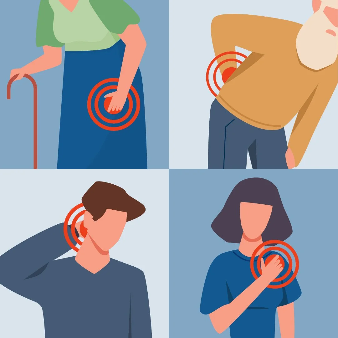
RAO provides joint pain injections at our Medical Imaging Center and our TimberRidge location, performed by our experienced team of Board-certified Radiologists.

Each year, millions of Americans are referred for a biopsy to analyze something that an initial exam or test reveals may be abnormal. A biopsy involves the removal of a small sample of affected tissue, whether via a needle, small incision or more-invasive excisional biopsy, depending on the area of concern.
WOmen’s Imaging Center (WIC)
News and Views Blog
Dual-energy x-ray absorptiometry (DEXA) is an imaging procedure used to measure the density of your bones. Bone densitometry testing is predominantly performed to assess the risk of osteoporosis, a condition characterized by low bone density, which produces an increased risk of fracture.
For the latest, most advanced mammography technologies, highly experienced doctors, skilled technologists, and a dedicated care team, the name to know is RAO.
A breast aspiration is one of the most common methods used to assess a fluid-filled cyst within the breast. During this procedure, a small amount of fluid or breast tissue is removed and examined to see if cancer cells are present. While about a vast majority of breast cysts are not cancerous, breast aspirations provide the information needed to determine the next best steps.

May 8–14 celebrates National Women's Health Week, highlighting the many ways that women can honor and take steps to protect their best health, vitality, energy and longevity.
If you need a biopsy, you have a choice of providers. Trust RAO’s 50 years serving Central Florida for experienced, dedicated and individualized care.
DEXA can help discover osteoporosis early, so treatments and lifestyle changes can be employed to help prevent fracture from happening.
Women’s Imaging encompasses a range of diagnostic and interventional imaging tests and services designed to meet the specific health needs of women. As technologies have improved, so have screening, diagnostic and treatment modalities for a host of diseases and disorders. Of foremost interest and importance to most women are the following …
FDA-approved breast tomosynthesis, also called 3D mammography, is a huge leap forward in both early breast cancer discovery and a reduction in stressful false positive results.

A local leader in the crucial area of breast biopsy, RAO has upgraded our suite of services to include the Brevera® breast biopsy system with CorLumina® imaging technology and Affirm® prone access.

Caught early, breast cancer has a nearly 100% five-year survival rate. RAO’s team of breast health specialists is dedicated to helping you flourish.

Years ago, breast biopsy involved surgery. Today, imaging technologies like 3D x-ray, ultrasound and MRI provide pinpoint guidance and quick, easy sampling on an outpatient basis.

Women at elevated risk of breast cancer can benefit from adding radiation-free Breast MRI to their regular mammography screening, for the earliest chance of detection.

3D is the only mammography system FDA-approved as superior for the 47% of women with dense breast tissue, which is difficult to image using 2D mammography.


Women who had a screening mammogram reveal something suspicious or are considered at high risk of breast cancer can benefit from the accuracy and detail of radiation-free breast MRI.

RAO is delighted to announce that breast imaging specialist Dr. Ridgely Meyers has joined their Women’s Imaging Center diagnostic team. Dr. Meyers has exceptional training and a personal commitment to the early discovery and treatment of breast cancer.

Since 1973, RAO has been dedicated to promoting and protecting health and quality of life through preventative screening exams and next-level diagnostic testing. Because women have specific healthcare needs, RAO established Women’s Imaging Center (WIC), the region’s first and only comprehensive, full-service center dedicated to women’s diagnostic services.

Uterine fibroids are the most common benign tumors in women of childbearing age, and can come with a host of symptoms, including heavy or painful periods, bleeding between periods, frequent urination and pain in the abdomen, back and/or during intercourse.

For decades, wire localization has been the preferred method for radiologists to mark a non-palpable tumor identified on a mammogram and/or breast ultrasound. In this procedure, wire markers are used to guide a surgeon to the tumor so they may perform breast-conserving therapy (BCT) such as a lumpectomy to remove a tumor while preserving healthy breast tissue, instead of removing the entire breast (mastectomy).

As the region’s only comprehensive women’s diagnostic services provider, RAO has replaced all 2D mammography equipment with FDA-approved 3D mammography, also known as tomosynthesis. Women naturally have questions about the difference, and we’re happy to explain the superiority of 3D mammography so you can feel confident about scheduling your routine breast cancer screening.

RAO’s Women’s Imaging Center (WIC) and TimberRidge Imaging Center want to remind women to maintain a “Rosie” outlook on a riveting subject: early breast cancer detection. Most people know of the importance of finding breast cancer early, when it is most easily and successfully treated. But many don’t know about some of the more sophisticated technologies that arm our dedicated faction of breast imaging subspecialists.

Breast MRI is a supplemental exam designed to deliver additional information about breast tissue and abnormal growths, and is particularly useful in helping your radiologist and referring clinician see through dense breast tissue.

Once you delve deeply into local practices, you’ll find that RAO’s Women’s Imaging Center (WIC) is the only comprehensive, full-service center dedicated to women’s diagnostic services.


No matter who you are, breast cancer can impact your life. Whether it affects you or a loved one, it can be a shocking diagnosis, even for those with inside knowledge of how common it is, and how important the need for early detection. Just ask Yvonne and Michele, two beloved members of RAO’s very own healthcare team, who both recently discovered they had breast cancer.

RAO is proud to have a dedicated team of superheroes in the battle against breast cancer, led by RAO’s Medical Director of the Women’s Imaging Center, Board Certified radiologist, Dr. Amanda Aulls.

The differences between 2D and 3D mammography™ exams are notable, especially for the estimated 47% of women with dense breast tissue. Dense breast tissue is closely-compacted, creating areas that can be nearly or completely invisible to radiation, making it difficult for 2D mammography to capture images of lesions, especially when they are small.

RAO is proud to have Board Certified radiologist Dr. Amanda Aulls as its Medical Director of Women’s Imaging Services at its Women’s Imaging Center and TimberRidge Imaging Center.
News
News and Views Blog


ITN recently spoke with Lisa Adams, CEO of Radiology Imaging Associates on the increase of women in radiology leadership roles. Read the interview here.

At some point or other, nearly everyone will need a radiological exam, but many people aren’t aware of what role the radiologist plays in their care.

AI’s capacity for deep learning and extraordinary speed is changing the landscape for radiologists, diagnostic testing and patient outcomes.

As a longtime forerunner in radiological technologies, RAO is committed to providing patients and referring clinicians with the fastest, safest and most accurate imaging systems available. For people needing rapid and reliable PET/CT imaging, RAO is proud to include the groundbreaking Biograph mCT.

The U.S. Preventive Services Task Force has updated its recommendations for low-dose computed tomography screening to include more people and bridge the gap in care among high-risk groups omitted from the previous criteria. Read more about the current recommendations.
A local leader in the crucial area of breast biopsy, RAO has upgraded our suite of services to include the Brevera® breast biopsy system with CorLumina® imaging technology and Affirm® prone access.

April 26 – 30 is Patient Experience Week, a time for RAO and practices across the nation to celebrate, honor and express gratitude to patients and the hardworking healthcare teams dedicated to providing outstanding and heartfelt care.
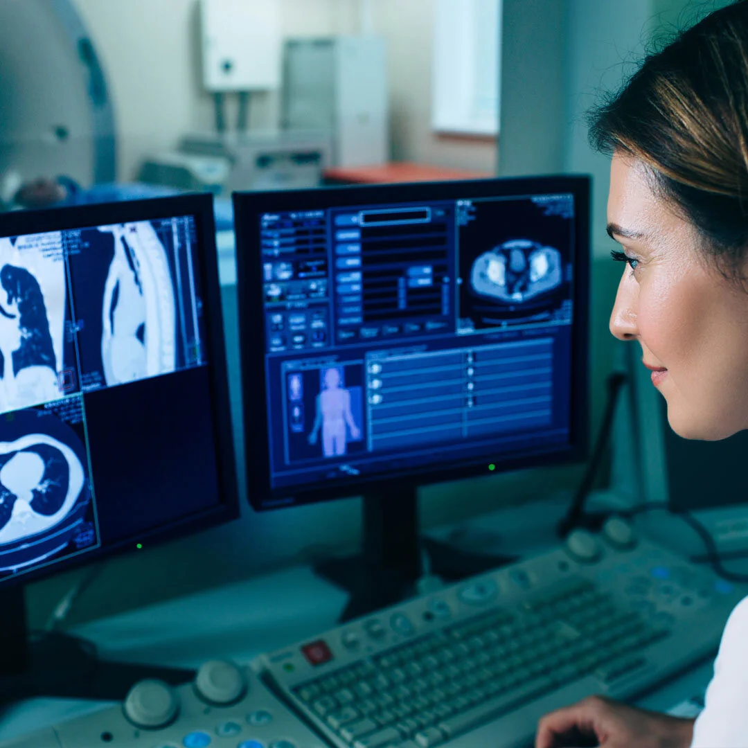
To continue providing important screening and diagnostic services while reducing the risk of COVID-19, many radiology practices, including RAO, have expanded their current level of teleradiology service, reading applicable images remotely.

Don’t let fear or anxiety take your life off-course. The sooner you get your appointment scheduled and over with, the sooner you can get back to your best possible life.

RAO is delighted to announce that breast imaging specialist Dr. Ridgely Meyers has joined their Women’s Imaging Center diagnostic team. Dr. Meyers has exceptional training and a personal commitment to the early discovery and treatment of breast cancer.

February is American Heart Month. During a time when many people are focused on Valentine’s hearts, it’s also a time to take stock of the muscle that keeps us alive and kicking.

As part of an ongoing effort to provide patients and referring clinicians with the quickest and most accurate diagnoses and services, RAO has added five new radiologists to our care team, boosting the number of doctors to 23.

November is Lung Cancer Awareness month, and for good reason: lung cancer is the number one cause of cancer death in the US. While lung cancer has multiple causes, about 85% of cases are associated with long-term tobacco smoking.

For most people, hearing the word “biopsy” is unsettling. Luckily, most biopsies turn out to be negative, meaning there are no signs of cancer. Infection, internal injury and other diseases can create similar symptoms, so when your doctor or clinician orders a biopsy, it is important to get checked out as quickly as possible.

The Internet can be a great resource for nearly everything, but incorrect, even false, information abounds, especially related to health and wellness.


RAO is pleased to announce the addition of interventional radiologist Dr. Ray Sutkowski to our Center for Vascular Health team.

Low-Dose Computed Tomography, or LDCT, is an annual screening exam that can catch lung cancer while it is still tiny and localized, before it causes symptoms.

RAO is always looking for ways to expand and enhance our care to patients, and that includes introducing accomplished new members to our service team. Edward Todd, PA-C, is a Physician Assistant certified by the National Commission of Certification of PAs….

Patients of Seven Rivers Medical Center in Citrus County will benefit from enhanced diagnostic and interventional radiology services, thanks to a new partnership with Radiology Associates of Ocala
RAO is proud to have Board Certified radiologist Dr. Amanda Aulls as its Medical Director of Women’s Imaging Services at its Women’s Imaging Center and TimberRidge Imaging Center.

“I’m proud to be part of the RAO team and grateful to have the opportunity to bring my experience to the local community.”
Center for Vascular Health (CVH)
News and Views Blog


Hearts are strengthened by commonsense habits, some of which you have likely heard before. But reminders never hurt, especially not ones this vital. Adopting better habits today can make all the difference in your heart health going forward.

If you suffer from varicose veins, don’t let them impact the way your legs look and feel another day. Interventional Radiology can provide safe varicose vein treatment with less risk, quicker recovery and minimal scarring.

CVH’s board-certified Interventional Radiologists deliver an array of services addressing vascular and nonvascular issues from blocked arteries to varicose veins to pain injections, with minimal invasion and risk.

Vascular and Interventional Radiologist Dr. Jaime Abesada shares his thoughts on new technologies in the field and his hopes for the future of patient care.

Dr. Kareem Bohsali offers his views on radiology’s ever-growing role in the diagnosis and treatment of disease.
Uterine fibroids are the most common benign tumors in women of childbearing age, and can come with a host of symptoms, including heavy or painful periods, bleeding between periods, frequent urination and pain in the abdomen, back and/or during intercourse.

Peripheral vascular disease, or PVD, is a broad term that covers a wide scope of disorders affecting the blood vessels. Most often, PVD is caused by peripheral artery disease, which is a hardening of the arteries known as atherosclerosis.

Endovenous Laser Ablation (ELA) is an image-guided procedure that uses laser-generated heat to close off and shrink varicose veins. Over the past 10-15 years ELA has proven to be so safe and effective that it has replaced most invasive surgical techniques.

Phlebectomy is a minimally invasive procedure used to remove varicose veins, and is the preferred method of treatment for some patients. It is typically performed using local anesthesia to numb the skin above the vein, so your interventional radiologist can make a very small incision without causing discomfort.

Many people suffering from liver cancer and related conditions now have a minimally invasive treatment option called transarterial chemoembolization, or TACE. TACE can be used to treat primary or metastasized liver cancer (cancer that has spread to the liver from another organ), as well as cancer of the bile ducts of the liver, a disease called cholangiocarcinoma.

RAO’s Center for Vascular Health (CVH) provides a gamut of next-generation vascular and non-vascular care and a team of experienced and compassionate interventional radiologists dedicated to your health, safety and comfort.
February is American Heart Month. During a time when many people are focused on Valentine’s hearts, it’s also a time to take stock of the muscle that keeps us alive and kicking.
RAO is pleased to announce the addition of interventional radiologist Dr. Ray Sutkowski to our Center for Vascular Health team.
RAO is always looking for ways to expand and enhance our care to patients, and that includes introducing accomplished new members to our service team. Edward Todd, PA-C, is a Physician Assistant certified by the National Commission of Certification of PAs….

If you suffer from varicose veins, you owe it to yourself to find out how quickly and easily they can become a thing of the past.
Community
News and Views Blog
ITN recently spoke with Lisa Adams, CEO of Radiology Imaging Associates on the increase of women in radiology leadership roles. Read the interview here.
April 26 – 30 is Patient Experience Week, a time for RAO and practices across the nation to celebrate, honor and express gratitude to patients and the hardworking healthcare teams dedicated to providing outstanding and heartfelt care.
Wellness
News and Views Blog
With so many imaging centers dotted around Central Florida, it’s easy to surmise that medical imaging plays a major role in modern healthcare. But what is its specific function in the treatment of cancer? Actually, medical imaging is a primary component across every aspect of successful cancer treatment, from cancer discovery to ongoing analysis of disease progression to the monitoring of therapies.
Dual-energy x-ray absorptiometry (DEXA) is an imaging procedure used to measure the density of your bones. Bone densitometry testing is predominantly performed to assess the risk of osteoporosis, a condition characterized by low bone density, which produces an increased risk of fracture.
RAO offers gastric emptying scans at our Medical Imaging Center at Windsor Oaks. This facility is accredited in Nuclear Medicine by the American College of Radiology for safety, accuracy and best practice standards, so you know you’re receiving the highest level of care.
The American Cancer Society says that one in seven men will be diagnosed with prostate cancer during his lifetime. Caught and treated while it still localized, prostate cancer has a nearly 100% survival rate.
May 8–14 celebrates National Women's Health Week, highlighting the many ways that women can honor and take steps to protect their best health, vitality, energy and longevity.
Diagnostic ultrasound is an imaging exam that utilizes sound waves and computer technology to construct images of organs, structures, tissues and blood flow.
RAO offers the latest, most sophisticated gallbladder imaging, including ultrasound, MRI, CT and nuclear medicine scanning.
Because the parathyroid glands are tiny, they can be difficult to image, demanding a highly skilled and experienced radiologist or endocrine physician to promote the utmost level of accuracy.
Women’s Imaging encompasses a range of diagnostic and interventional imaging tests and services designed to meet the specific health needs of women. As technologies have improved, so have screening, diagnostic and treatment modalities for a host of diseases and disorders. Of foremost interest and importance to most women are the following …

Musculoskeletal imaging, or MSK, provides valuable information about the type, location and extent of a sports injury. RAO’s team of fellowship-trained radiologists specializes in MSK, meaning our doctors have additional years of training specifically in sports-related injuries, to accurately diagnose, assess, and, in some cases, assist in the treatment staging of fractures, sprains, tears, pulls, inflammation, overuse injuries and other issues.